Pink-violet diamonds from the Lomonosov mine: morphology, spectroscopy, nature of colour
Abstract
The article presents the results of the first comprehensive study of mineralogical and spectroscopic (IR, PL, EPR) characteristics of diamonds from the Lomonosov mine (Arkhangelskaya pipe) with a unique pink, pink-violet colour. It is shown that all crystals belong to the IaA type, with a total nitrogen content in the range of 500-1500 ppm, with a low degree of aggregation. The colour is heterogeneous, concentrated in narrow twin layers. It is presumably caused by the previously described M2 centres. The colour shade is affected by the content of P1 paramagnetic centres (C-defect). A positive correlation is observed between the colour saturation and the intensity of W7 paramagnetic centres. A convergent model of the formation of pink diamonds is assumed, according to which the determining factors are the ratio and concentration of structural impurities in the diamond, its thermal history, and conditions of plastic deformation, and not the origin of the diamond and the petrochemical properties of its host rocks.
Introduction
Diamonds in Fancy Pink and Fancy Purple-Pink colours are among the most expensive ones. The combination of a pink shade or colour with gem quality is so rare that it makes such diamonds unique. There are only six mines in the world with small but consistent finds of pink and purple diamonds. In Russia, pink and pink-violet diamonds are found in the Lomonosov mine and in the Internationalnaya pipe. Previously, using the examples of mines in Australia (Argyle), India, and Tanzania, several mechanisms for the formation of pink and purple colours were proposed, but the conditions for their occurrence have not yet been determined.
Several types of pink diamond colour are known [1-3]. They differ in their absorption spectra [4, 5] – either a broad structureless band in the region of 550 nm, or absorption systems with zero-phonon lines at 575 and 637 nm [6, 7]. The absorption systems at 575 and 637 nm belong to the nitrogen-vacancy centres NV0 and NV–, respectively [3, 8]. The pink colour of the first type is heterogeneous. In crystals it is localized in individual lamellae induced by plastic deformation [9-11]. The pink colour of the second type is less intense, but homogeneous. This nature of colour was found in some historical diamonds of India. Diamonds of this type have very low nitrogen concentrations [2, 4, 12]. In nature, such crystals are extremely rare, but for synthetic stones a technique was developed for obtaining a pink colour, associated specifically with the NV0 and NV– centres. On the contrary, the absorption band at 550 nm cannot yet be reproduced when growing artificial diamonds [13].
Pink diamonds due to the absorption band with a maximum of 550 nm, in turn, are divided into two types, differing in morphological features, spectroscopic characteristics, and association with certain mines [3, 9]. Their main difference is the concentration of nitrogen in Ntot and the degree of its aggregation NB, i.e., the ratio of nitrogen in the form of B-centres NB (four nitrogen atoms and a carbon vacancy) to the total concentration of nitrogen – in the form of B and A-centres NA (two neighbouring nitrogen atoms in carbon positions of the crystal structure). The first type is known from diamonds from the Argyle pipe (Australia) [5, 11, 14], and similar diamonds also come from placer deposits in Venezuela [10]. The total nitrogen content in them is usually low, to 250 ppm, and the degree of its aggregation is high, the NB/NA concentration ratio is greater than unity (IaA < B), often multiple. The colour is distributed unevenly throughout the crystals; it is mainly concentrated in layers – lamellas, often wavy, as well as in spots [13, 15]. There is no consensus among researchers about the mechanism of the appearance of pink colour [4, 10, 16].
The second type of pink diamond (IaA >> B), the colour of which is also determined by the 550 nm absorption band, is found in mines of Russia [3, 7, 17] (Internationalnaya, Arkhangelskaya, Karpinsky-1 pipes), South Africa [12], and Canada [8]. The total nitrogen content in crystals of this type is from 300 to 1200 ppm or more, and the NB/NA ratio is less than unity [8, 14]. The source for the colour of these crystals is studied in much more detail than for diamonds from the Argyle pipe, and is described in detail in [6, 7]. The pink, often with a violet shade, colour is concentrated in thin layers along {111}, in a twin orientation with respect to the main volume of the crystal. The colour intensity correlates with the concentration of the paramagnetic centre M2 contained in these microtwin layers, which has a deformation nature. It is likely that the M2 centre appears in the visible range as a colour centre and is responsible for the emergence of the 550 nm absorption band [3, 16]. The centre is formed by two nonequivalent nitrogen atoms separated by three carbon atoms, with the nitrogen atoms located along the ⟨001⟩ direction, forming opposite vertices of a polyhedron, similar in configuration to an octahedron with reduced symmetry [6, 16].
Pink, pink-violet crystals were found in the Arkhangelskaya and Internationalnaya kimberlite pipes [3], which according to the petrochemical classification [17] belong to low-titanium type rocks (TiO2 < 1 %), and in the Argyle lamproite pipe. Based on a number of petrological and geochemical characteristics [18, 19], low-titanium kimberlites are close to lamproites. A similar formation trend is assumed – olivine lamproites of the Argyle pipe – Al-kimberlites of the Zolotitsky field – Al-kimberlites of the Internationalnaya pipe [8, 19, 20]. Many works associate the defect and impurity composition of diamonds [14, 21-23], habitus [17, 24], the external appearance of diamond crystals, and the conditions of their formation and occurrence in mantle rocks [25-27].
This work studied the morphological and spectroscopic characteristics of pink-violet diamonds from the Arkhangelskaya pipe of the Lomonosov mine. The purpose of the study was to determine the features of the external and internal structure of diamonds formed in the post-growth period as a result of mechanical and temperature influences, as well as to assess the composition of structural defects and their possible effect on the pink colour of the crystals.
Research materials and methods
Seven pink diamond crystals of various shades and saturations from batches of commercial mining of the Arkhangelskaya pipe in 2010-2020 were studied. (Fig.1, a). A mineralogical description was made, studies were carried out using infrared (IR) and photoluminescence (PL) spectroscopy, as well as electron paramagnetic resonance (EPR). The systematics of structural defects is given according to [17, 21, 25]. Sample 1784-8-1 was polished after spectroscopic studies, which made it possible to obtain an absorption spectrum in the visible region.
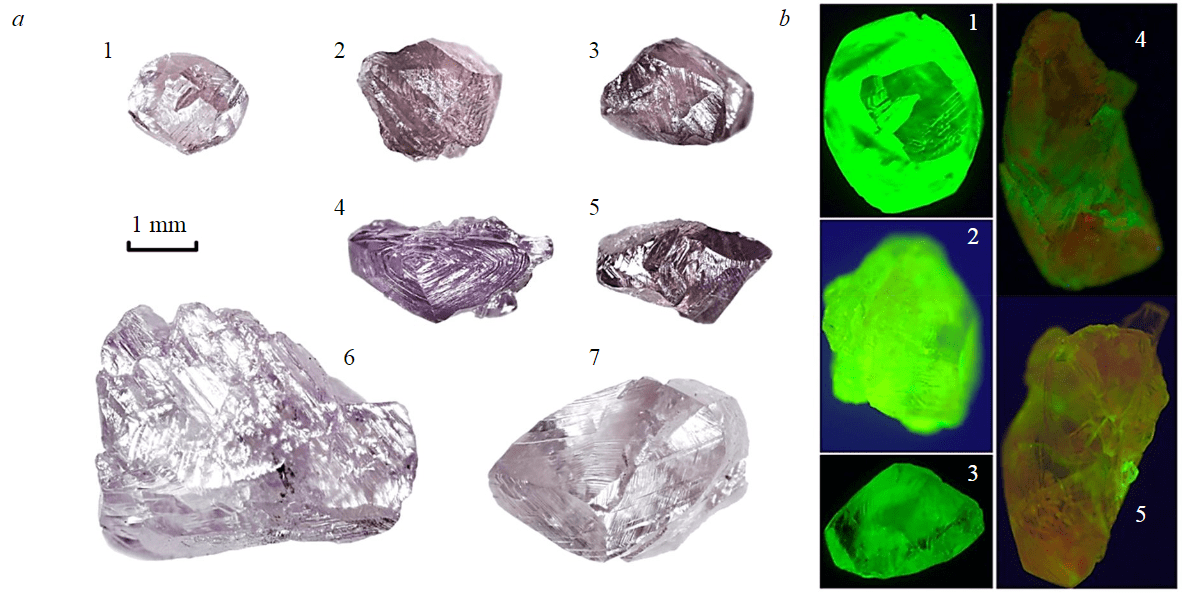
Fig.1. Studied diamond crystals from the Arkhangelskaya pipe: photos and images of photoluminescence of crystals when excited by a 405 nm laser 1 – sample 1784-8-1; 2 – sample 1784-41-2; 3 – sample 1784-41-6; 4 – sample 1784-41-11; 5 – sample 1784-41-14; 6 – sample A0058-1; 7 – sample 1797-3
The morphology of the crystals was studied under a Motic SMZ-143 binocular with a UV illuminator box (365 nm). The recording of IR spectra was carried out on a Vertex-70 Fourier spectrometer (Bruker) with a Hyperion-1000 microscope. The total nitrogen concentration (Ntot) and the fraction of nitrogen in the form of B defects (NB, %) were calculated using known proportionality coefficients. The lower limit for the detection of nitrogen in B form is 5-7 %. The B′ band with a maximum in the region of 1360 cm–1 also indicates the presence of B defects. Planar B' defects (platelets) are composed of interstitial carbon atoms, the concentration of which is proportional to the amplitude of the aВ′ band, and the position of the band maximum depends on the size [14]. The presence and relative content of hydrogen-containing centres was determined in accordance with [28].
The absorption spectrum in the visible region was obtained using a LOMO MSFU-K microscope-spectrophotometer. PL spectra were recorded on an InVia spectrometer (Renishaw) with excitation by lasers of 405, 488, and 785 nm with an objective (×5) at a temperature of 77 K [29].
EPR spectra were recorded on a CMS-8400 spectrometer in the X-band (~9.4 GHz) at a modulation amplitude of 0.1 mT, a modulation frequency of 100 kHz, and a microwave radiation power of 0.2 and 5 mW. The samples were adjusted in L4 || H according to the angular dependence of the P1 centre; shootings were made with significant accumulation (to 75 scans).
Research results
The studied samples are represented by dodecahedroids and crystals of combination shape with manifestations of octahedron faces. Pink crystals of octahedral habit in the Arkhangelskaya pipe are rare; their finds during the entire period of mining of the pipe are singular. The morphology of the crystal surface is specific. Its main feature is the strong influence of plastic deformation (Fig.1, a). All crystals are formed by a tangential growth mechanism and have a thin-layered structure, expressed in the surface morphology by a sharp concentric hatching around the L3 axes. Lamellae of plastic deformation are also observed in two or three crystallographic directions (Fig.2). Superimposed sharp shifts of lamellae of plastic deformation penetrate the entire volume of the crystal and are observed on the surface from opposite sides in the form of parallel stripes and steps, together with the main shading they form a large block topography. The resulting steps often go deeper into the gem volume. The surface of the blocks is matte (covered with fine shagreen) (Fig.3). According to these characteristics and the degree of their expression, pink crystals differ not only from colourless and yellow ones, but also from brown crystals, also subjected to plastic deformation. Almost all crystals have large protomagmatic and minor technogenic chips. When viewed in polarized light, areas with internal stresses and characterized by anomalous birefringence are clearly visible. Signs of linear colour dichroism appear when viewed in different orientations relative to the directions with the most pronounced lamellae of plastic deformation.
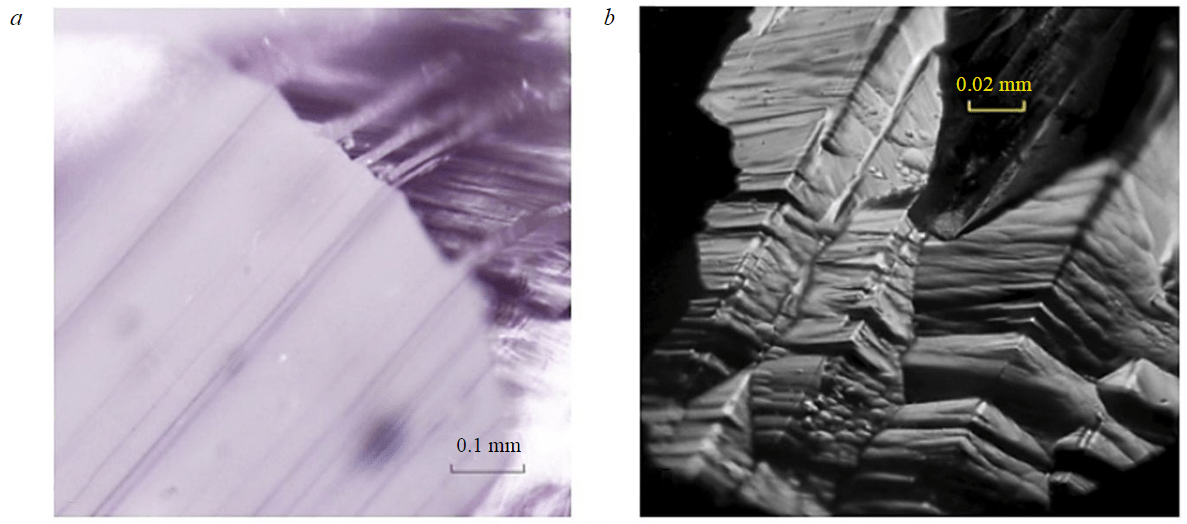
Fig.2. Plastic deformation planes in pink-violet diamond 1784-8-1: a – boundary of the ground area and the crystal surface (macrophotography); b – emergence of a plastic deformation band on the crystal surface (micrograph)
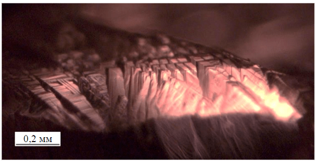
Fig.3. Matte surface (micrograph of sample A0058-1)
Visual photoluminescence (under a 365 nm UV lamp) of moderate intensity is manifested in yellowish or yellowish-bluish shades (see Fig.1, b). Low-intensity systems N3, 491 nm, H3 are observed in the PL spectra upon excitation with a 405 nm laser.
When excited by a 488 nm laser, an intense H3 system with a zero-phonon line at 503 nm, a peak at 612 nm, and a wide structureless band with a maximum at about 720 nm is recorded in the spectra of all samples. The relative intensities of these systems vary widely, both between crystals and within the volume of some samples. Peaks at 700, 787, 926 nm are recorded in the spectra of most crystals; in some samples – 523, 694 nm. If the 787 nm line is recorded in the spectrum excited by a 488 nm laser, then the phonon wing of this system determines the shape of the spectrum when excited by 785 nm, and the high intensity of the 926 nm line is noted. In samples 1797-3 and 1784-41-14, a system with maxima at 868, 895, 923, 952, 983, 1018 nm (energy shift 0.042 eV) is observed. In other samples this system is not observed, probably against the background of the phonon wing of the 787 and 926 nm systems. This set of PL features is typical for crystals with plastic deformation [29]. When excited at 785 nm, a system with a maximum at 986 nm (H2) is observed in the spectra of all crystals. The uneven volume distribution of luminescence in some crystals is shown in Fig.1, b. Crystals 1784-8-1, 1784-41-2, 1784-41-6 have uniform green PL of the H3 system, and in samples 1784-41-11 and 1784-41-14 there are regions with dominance of both red luminescence (720 nm band) and a brighter green one. The results of studies using IR, EPR, and PL spectroscopy methods are given in the Table.
In the IR absorption spectra of most samples, only the nitrogen A-defect and the 3107 cm–1 line with satellites, corresponding to the hydrogen-containing centre VN3H [28] (Fig.4, a), are revealed. In four samples, a band with a maximum at around 4160 cm–1 is observed; the Table shows its amplitude. This band is the “amber” centre; it is typical in the spectra of brown crystals with signs of plastic deformation. The IR absorption spectra of four crystals reveal additional peaks at 3311, 3189, 3154, 3144, 3050 cm–1; the amplitude of peak 3154 (a3154) is given in the table. This set is typical for a diamond without B-defects from the Arkhangelskaya and Karpinsky-I pipes [18]. All diamonds with a pink and violet-pink shade are medium-nitrogen and high-nitrogen with a very low degree of nitrogen aggregation into the B-defect (Fig.4, a, table). The B′ band was not detected in the spectra of two crystals. In the spectra of three crystals, it had an amplitude of less than 1 cm–1, which indicates a very low concentration of B-defects; they are not observed in the spectra.
Results of crystal studies using IR, EPR, and PL spectroscopy
|
Study |
Samples |
||||||
|
1784-8-1 |
1784-41-2 |
1784-41-6 |
1784-41-11 |
1784-41-14 |
A0058-1 |
1797-3 |
|
|
Diamond colour |
Pink Purple |
Pink Purple |
Pink Purple |
Purple |
Dark Purple |
Pink |
Light purple pink |
|
Weight, mg |
8 |
5,5 |
5,5 |
8 |
7 |
70 |
45 |
|
Ntot, ppm |
1146 |
850 |
990 |
1330 |
840 |
570 |
540 |
|
NA, ppm |
1015 |
800 |
990 |
1330 |
840 |
492 |
420 |
|
NB, ppm |
130 |
– |
– |
– |
– |
108 |
120 |
|
NB, % |
11 |
– |
– |
– |
– |
19 |
22 |
|
aB′/vB′, cm–1 |
0.3/1380 |
– |
– |
0.8/1382 |
0.5/1377 |
2.5/1365 |
4.6/1365 |
|
a3107, cm–1 |
7,0 |
10.8 |
13.5 |
6.1 |
6.5 |
0.15 |
0.12 |
|
a4160, cm–1 |
0.12 |
0.1 |
0.2 |
– |
0.41 |
– |
– |
|
a3154, cm–1 |
0.16 |
0.44 |
0.6 |
0.28 |
– |
– |
– |
|
EPR centres* |
P1, W7, N2, M2 |
W7, P1, N2, M2 |
W7, P1, N2, M2 |
W7, P1, N2, M2 |
W7, N2, P1, M2 |
P1, M2, M5 |
P1, N2, W7, M2 |
|
λех = 488 nm** |
Н3, 612, 720w***, 694, 700, 787, 926 |
Н3, 612, 720w, 694, 700, 787, 926 |
Н3, 612, 720w, 694, 700, 787, 926 |
Н3, 612, 720w, |
Н3, 612, 720w, 694, 700, 787, 926 |
Н3, 720w |
Н3, 612, 580w, 720w |
|
λех = 787 nm** |
926, 904, 986 |
926, 904, 986 |
926, 904, 986 |
986 |
926, 986 |
820/896/925 |
986 |
* A paramagnetic centre was identified that is dominant in intensity in the P1-W7 pair.
** The most intensive systems are in bold, and the least intensive systems are in italics.
*** 720w – wide band with a maximum at 720 nm, 580w – with a maximum at 580 nm.
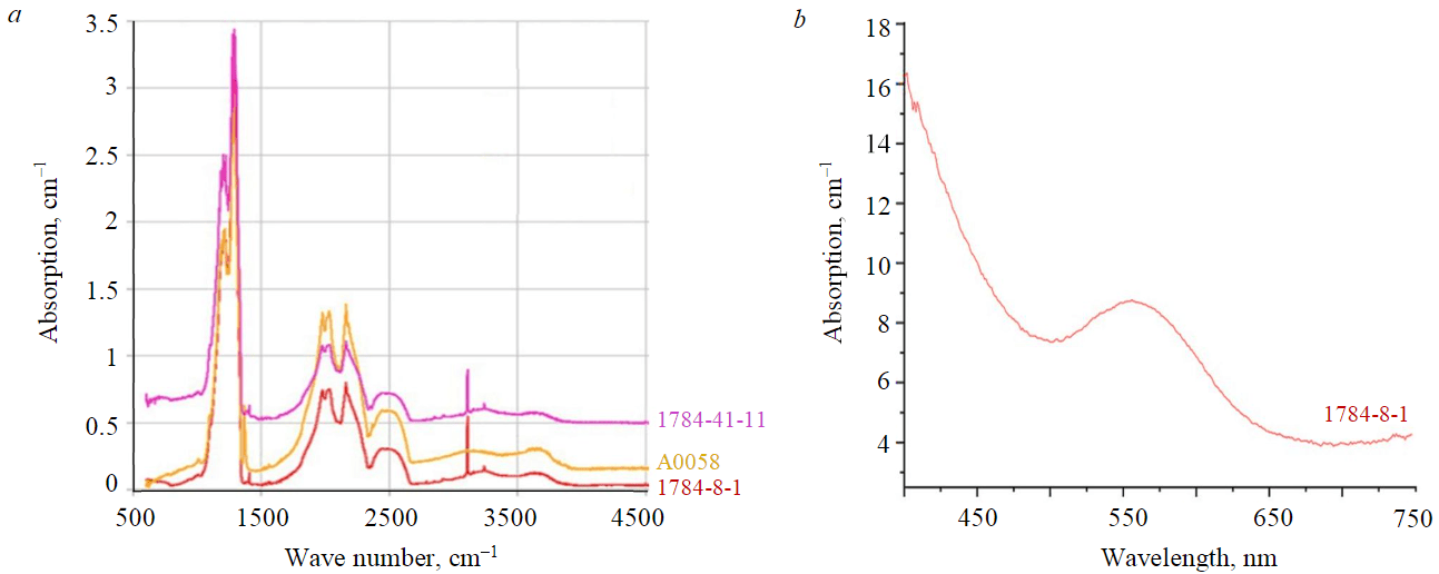
Fig.4. Absorption spectra of pink diamonds from the Arkhangelskaya pipe in the IR (a) and visible (b) regions
In the absorption spectrum in the visible region (Fig. 4, b), obtained after polishing sample 1784-8-1, a wide band appears in the region of 550 nm, which determines its pink colour, as well as a side absorption wing in the short-wave part of the spectrum, due to the presence of C-defects.
Discussion of the results
The pink colour in the studied samples is heterogeneous. It is concentrated in thin layers along {111}, coinciding in orientation with the bands of plastic deformation (see Fig.2). Areas with bending, “wavy” stripes of colour distribution, or with a “spotty” character described for crystals from the Argyle pipe [13], were not found in the studied samples. The layers of intense pink colour are confined to the lamellae of plastic deformation revealed in the topography, as can be seen in the example of polished sample 1784-8-1 (see Fig.2, a). The reason for this relationship is revealed with further magnification: visually observed bands of plastic deformation mark crystal areas with a microtwin structure (see Fig.2, b). The matte surface (fine shagreen) of some blocks is explained by the intersection of plastic deformation structures (see Fig.3).
Thus, the microstructure of the distribution of pink colour and the present violet shade in the studied diamond crystals corresponds to the colour model described in [3, 16], i.e., its concentration in thin plates in twin orientation [6, 10].
The results of the absorption study in the IR region are fully consistent with the identified colour distribution model. All samples have a nitrogen content of about 500-1500 ppm, which is significantly higher than the typical range for Argyle pipe diamonds (less than 250 ppm). At the same time, two pink samples (A0058-1, 1797-3) have a lower content of total nitrogen (700 and 540 ppm, respectively), and for more intensely coloured purple ones (2-5 in Fig.1, a) Ntot is in the range of 950-1500 ppm. This correlation is violated by the weakly coloured crystal 1784-8-1 (1 in Fig.1, a), for which Ntot = 1010 ppm. All studied diamonds belong to group IaA (A > B), the NB value is less than 22 %. A high total nitrogen content (NA + NB), a low degree of aggregation of centres into the B-form, and an increased concentration of C-defects (N+) in crystals of the octahedron – dodecahedroid series are typical for most colourless and yellow diamonds from the Lomonosov mine [17, 21]. For most samples, high relative contents of hydrogen-containing VN3H centres were noted.
M2 centres, formed as a result of plastic deformation and presumably being centres of pink colour, were found in all samples. Paramagnetic centres P1 were also found in all samples (Fig.5, Table) [16]. The paramagnetic centre P1 is a single substituting nitrogen atom (C-defect). Due to the low concentration, C-defects in the studied diamonds are detected only by EPR. C system (main peak at 1130 cm–1) does not appear in the IR absorption spectra. In the diamonds of the Lomonosov mine C-defects affect the perception of violet and pink colours: absorption in the short-wave part of the spectrum reduces the proportion of cold tones in the colour balance, shifting the resulting colour closer to warm pink. At first glance, additional absorption in the short-wave part of the visible region (see Fig.4, b) should make the crystal darker, but the opposite effect is observed. Samples 1784-8-1, A0058-1, and 1797-3, in which the EPR centre P1 (C-defect) is most pronounced, are lighter (see Table). This can be seen in Fig.1, a.
In the table, the paramagnetic centres that dominate in intensity in the P1-W7 pair are in bold. The W7 centre is a ring consisting of four carbon atoms and two nitrogen atoms located in opposite areas. According to modern concepts, W7 centres are formed from A centres during plastic deformation of diamond crystals, with a local decrease in the symmetry of the crystal, being one of the main markers of this process [3, 16]. Based on the fact that the M2 centres also have a deformation nature [16], it seems logical to have a positive correlation between the intensity of the lines of the W7 centre as a marker of plastic deformation, and the saturation of the pink-violet colour. However, it should be taken into account that W7 centres are formed in a much wider temperature range compared to M2 centres and are found much more often in different types of diamonds. The above-mentioned division of the studied crystals according to colour intensity fully corresponds to the compositional features of the paramagnetic centres in them. For example, EPR spectrum of sample A0058-1 (Fig. 5, a) has a noticeably higher intensity of the lines of the P1 centres (single substitutions of carbon with nitrogen) compared to the lines of the W7 centre, its colour is lighter and more pink. The opposite correlation, for example, for crystal 1784-41-14 (Fig.5, b), leads to a more saturated violet colour.
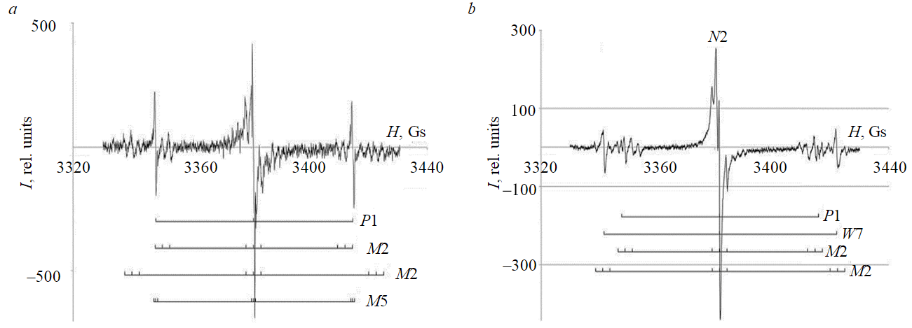
Fig.5. EPR spectra of the studied diamonds: a – pale pink crystal A0058-1; b – more intensely coloured crystal 1784-41-14
Also, crystals with C-defects are characterized by the H2 PL system [1, 12], which was found in all samples. The H2 defect is an ionized H3 centre [12]. Apparently, the H2 defect arises from the interaction of two NV centres, which are characterized by high mobility [6]. Previously, the presence of C-defects in pink diamond was revealed when studying the photochromism of crystals with a high proportion of B-defects [1, 5]. One of the sources of C-defects is the destruction of B-defects during plastic deformation, with the formation of C, H3, N3 defects [12, 18, 30]. Thus, both pink crystals without B defects and crystals with a high proportion of nitrogen in the B form (from the Argyle mine) have C defects that contribute to the perception of pink colour.
The ratio and concentration of nitrogen and hydrogen centres in diamonds from the Lomonosov mine are close to those in purple diamonds from the Internationalnaya kimberlite pipe. The 550 nm absorption band is the reason for the pink colour not only in the studied diamonds of the Arkhangelskaya kimberlite pipe, but also in the diamonds of the Argyle lamproite pipe, which differ from them in many characteristics [2, 5, 8]. It should also be noted that there is a similarity in the uneven distribution of colour caused by the 550 nm band for diamond crystals from both kimberlite and lamproite deposits. A comparison of this factual material allows us to assume about the convergent nature of pink diamonds, which depends to a much greater extent on the nature of plastic deformation and the PT conditions of its occurrence than on the origin of the diamond and the properties of its host rocks. At the same time, an unambiguous sign of the petrochemical similarity of the mentioned diamond pipes – low TiO2 content – becomes understandable in the light of [19, 20], where low titanium content is considered as an indicator of high pressure during the kimberlites formation. Most kimberlite bodies in the Yakutia diamond province have a moderate content of titanium oxide (TiO2 1-2.5 wt.%) and large depths of origin of both diamonds [20, 24, 31] and proto-kimberlite magmas [25, 32, 33], therefore, higher temperatures of post-crystallization annealing [24, 34, 35], including during the movement of diamond-bearing rocks to the surface. High temperature and lower pressure contributed to the plastic deformation through the mechanism of dislocation sliding with the formation of brown diamonds, characteristic of 25-50 % of crystals from the Udachnaya, Zapolyarnaya, Komsomolskaya kimberlite pipes [24, 25, 31].
Thus, the observed empirical pattern of diamonds distribution according to the presence of pink colour with TiO2 content in diamond-bearing rocks is a consequence of the influence of the main factor – pressure and temperature conditions. An example of the influence of PT conditions is the Lomonosov mine (Arkhangelskaya and Karpinsky-1 pipes), which belongs to the low-titanium type of kimberlites and contains pink-violet diamonds, and the Grib mine, which belongs to the moderate-titanium type of kimberlites [19] and does not contain pink and purple diamonds, despite the spatial, geological, and temporal proximity of these mines [36-38], as well as the general similarity of mineralogy and defect and impurity composition of diamonds [17, 39].
Conclusion
The studied pink and pink-violet diamonds from the Lomonosov mine are medium- and high-nitrogen, with a low degree of aggregation of nitrogen centres and belong to the IaA > B group. In all crystals, P1 (C) and M2 centres are identified by the EPR, and nitrogen-vacancy systems H3 and H2 are identified from the luminescence spectra. The pink colour is heterogeneous, concentrated in thin layers along {111}, coinciding with the lamellae of plastic deformation of the crystals. M2 centres, presumably pink colour centres, were found in all samples. A positive correlation was observed between the intensity of the lines of the W7 paramagnetic centre as a marker of plastic deformation and the saturation of the pink-violet colour. The colour shade is associated with the intensity balance in the P1-W7 pair of paramagnetic centres. The presence of P1 centres (C-defects) causes absorption in the short-wave part of the visible region, giving the colour a warm shade. Violet, cold-shaded crystals are characterized by the dominance of the W7 paramagnetic centre.
The formation of pink and pink-violet diamonds is of a convergent nature: the presumed M2 colour centres are concentrated in microtwin lamellas, the formation of which requires certain PT conditions for the plastic deformation (relatively low temperatures and directed pressure). The appearance of pink diamonds in a particular mine depends to a greater extent on the presence of these conditions than on the origin of the diamond and the properties of its host rocks.
References
- Byrne K.S., Chapman J.G., Luiten A.N. Photochromic charge transfer processes in natural pink and brown diamond. Journal of Physics: Condensed Matter. 2013. Vol. 26. N 3. N 03551. DOI: 10.1088/0953-8984/26/3/035501
- Eaton-Magaña S., McElhenny G., Breeding C.M., Ardon T. Comparison of gemological and spectroscopic features in type IIa and Ia natural pink diamonds. Diamond and Related Materials. 2020. Vol. 105. N 107784. DOI: 10.1016/j.diamond.2020.107784
- Titkov S.V., Shigley J.E., Breeding C.M. et al. Natural-Colour Purple Diamonds from Siberia. Gems and Gemology. 2008. Vol. 44. N 1, p. 56-64. DOI: 10.5741/GEMS.44.1.56
- Gaillou E., Post J.E., Bassim N.D. et al. Spectroscopic and microscopic characterizations of colour lamellae in natural pink diamonds. Diamond and Related Materials. 2010. Vol. 19. Iss. 10, p. 1207-1220. DOI: 10.1016/j.diamond.2010.06.015
- Byrne K.S., Anstie J.D., Chapman J., Luiten A.N. Infrared microspectroscopy of natural Argyle pink diamond. Diamond and Related Materials. 2012. Vol. 23, p. 125-129. DOI: 10.1016/j.diamond.2012.01.032
- Titkov S.V., Krivovichev S.V., Organova N.I. Plastic deformation of natural diamonds by twinning: evidence from X-ray diffraction studies. Mineralogical Magazine. 2012. Vol. 76. Iss. 1, p. 143-149. DOI: 10.1180/minmag.2012.076.1.143
- Yuryeva O.P., Rakhmanova M.I., Zedgenizov D.A., Kalinina V.V. Spectroscopic evidence of the origin of brown and pink diamonds family from Internatsionalnaya kimberlite pipe (Siberian craton). Physics and Chemistry of Minerals. 2020. Vol. 47. Iss. 20. DOI: 10.1007/s00269-020-01088-5
- King J., Shigley J.E., Jannucci C. Exceptional Pink to Red Diamonds: A Celebration of the 30th Argyle Diamond Tender. Gems and Gemology. 2014. Vol. 50. N 4, p. 268-279. DOI: 10.5741/GEMS.50.4.268
- Gaillou E., Post J.E., Rose T., Butler J.E. Cathodoluminescence of Natural, Plastically Deformed Pink Diamonds. Microscopy and Microanalysis. 2012. Vol. 18. Iss. 6, p. 1292-1302. DOI: 10.1017/S1431927612013542
- Howell D., Fisher D., Piazolo S. et al. Pink colour in Type I diamonds: Is deformation twinning the cause? American Mineralogist. 2015. Vol. 100. N 7, p. 1518-1527. DOI: 10.2138/am-2015-5044
- Shigley J.E., Chapman J., Ellison R.K. Discovery and Mining of the Argyle Diamond Deposit, Australia. Gems and Gemology. 2001. Vol. 37. N 1, p. 26-41. DOI: 10.5741/GEMS.37.1.26
- Smit K.V., D’Haenens-Johansson U.F.S., Howell D. et al. Deformation-related spectroscopic features in natural Type Ib-IaA diamonds from Zimmi (West African craton). Mineralogy and Petrology. 2018. Vol. 112. Iss. 1, p. 243-257. DOI: 10.1007/s00710-018-0587-6
- Eaton-Magaña S., Ardon T., Smit K.V. et al. Natural-Colour Pink, Purple, Red, and Brown Diamonds: Band of Many Colours. Gems and Gemology. Vol. 54. N 4, p. 352-377. DOI: 10.5741/GEMS.54.2.352
- Taylor W.R., Jaques A.L., Ridd M. Nitrogen-defect aggregation characteristics of some Australasian diamonds: Time-temperature constraints on the source regions of pipe and alluvial diamonds. American Mineralogist. 1990. Vol. 75, p. 1290-1310.
- 15.Levashova E.V., Skublov S.G., Popov V.A.Distribution of Trace Elements Controlled by Sector and Growth Zonings in Zircon from Feldspathic Pegmatites (Ilmen Mountains, the Southern Urals). Geosciences. 2021. Vol. 7, p. 1-21. DOI: 10.3390/geosciences11010007
- Gavrilchik A.K., Skublov S.G., Kotova E.L. Rare element composition of beryl from the Sherlovaya Gora deposit, South-Eastern Transbaikalia. Zapiski Rossiiskogo mineralogicheskogo obshchestva. 2021. Vol. 150. N 2, p. 69-82. DOI: 10.31857/S0869605521020052
- Garanin V., Garanin K., Kriulina G., Samosorov G. Diamonds from the Arkhangelsk Province, NW Russia. Switzerland: Springer, 2021. 248 p. DOI: 10.1007/978-3-030-35717-7
- Melnik A.E., Korolev N.M., Skublov S.G. et al. Zircon in mantle eclogite xenoliths: a review. Geological Magazine. 2021. Vol. 158. Iss. 8, p. 1371-1382. DOI: 10.1017/S0016756820001387
- Kononova V.A., Bogatikov O.A., Kondrashov I.A. Kimberlites and Lamproites: Criteria for Similarity and Differences. Petrology. 2011. Vol. 19. N 1, p. 34-54. DOI: 10.1134/S0869591111010024
- Voitekhovskii Yu.L. Once again about the Curie dissymmetry principle. Zapiski Rossiiskogo mineralogicheskogo obshchestva. 2019. Vol. 148. N 3, p. 118-129 (in Russin). DOI: 10.30695/zrmo/2019.1483.09
- Pashkevich M.A., Alekseenko A.V. Reutilization prospects of diamond clay tailings at the Lomonosov mine, Northwestern Russia. Minerals. 2020. Vol. 10, p. 517-517. DOI: 10.3390/min10060517
- Harris J.W., Smit K.V., Fedortchouk Y., Moore M. Morphology of Monocrystalline Diamond and its Inclusions. Reviews in Mineralogy and Geochemistry. 2022. Vol. 88. Iss. 1, p. 119-166. DOI: 10.2138/rmg.2022.88.02
- Voitekhovskii Yu.L. Dodecahedral-icosahedral system. Zapiski Rossiiskogo mineralogicheskogo obshchestva. 2020. Vol.149. N 6, p. 101-109 (in Russian). DOI: 10.31857/S0869605520060155
- Zinchuk N.N., Koptil V.I. Typomorphism of diamonds of the Siberian Platform. Moscow: ООО “Nedra-Biznestsentr”, 2003, p. 603 (in Russian).
- Kostrovitskii S.I., Spetsius Z.V., Yakovlev D.A. et al. Atlas of primary diamond deposits in the Yakutia kimberlite province. Mirnyi: ALROSA, 2015, p. 480 (in Russian).
- Fedortchouk Y., Liebske C., McCammon C. Diamond destruction and growth during mantle metasomatism: An experimental study of diamond resorption features. Earth and Planetary Science Letters. 2019. Vol. 506, p. 493-506. DOI: 10.1016/j.epsl.2018.11.025
- Stachel T., Cartigny P., Chacko T., Pearson D.G. Carbon and Nitrogen in Mantle-Derived Diamonds. Reviews in Mineralogy and Geochemistry. 2022. Vol. 88. Iss. 1, p. 809-875. DOI: 10.2138/rmg.2022.88.15
- Goss J.P., Briddon P.R., Hill V. et al. Identification of the structure of the 3107 cm−1 H-related defect in diamond. Journal of Physics: Condensed Matter. 2014. Vol. 26. N 145801. DOI: 10.1088/0953-8984/26/14/145801
- Vasilev E.A. Luminescence of plastically deformed diamond in the range 800-1050 nm. Journal of Applied Spectroscopy. 2019. Vol. 86. N 3, p. 512-515. DOI: 10.1007/s10812-019-00850-0
- Nadolinnyi V.A., Yureva O.P., Eliseev A.P. et al. Destruction of nitrogen B1 centres during plastic deformation of natural diamonds of 1aB type and the behaviour of the resulting defects during PT treatment. Doklady Akademii nauk. 2004. Vol. 399. N 4, p.532-536 (in Russian).
- Vyatkin S.V., Kriulina G.Yu., Garanin V.K. et al. Morphology and defect and impurity composition of diamonds from the Zapolyarnaya pipe. Vestnik Moskovskogo universiteta. Seriya 4. Geologiya. 2021. N 1, p. 99-109 (in Russian). DOI: 10.33623/0579-9406-2021-1-99-109
- Serebryakov E.V., Gladkov A.S. Geological and structural characteristics of deep-level rock mass of the Udachnaya pipe deposit. Journal of Mining Institute. 2021. Vol. 250, p. 512-525. DOI: 10.31897/PMI.2021.4.4
- Duryagina A.M., Talovina I.V., Lieberwirth H., Ilalova R.K. Morphometric parameters of sulphide ores as a basis for selective ore dressing. Journal of Mining Institute. 2022. Vol. 256, p. 527-538. DOI: 10.31897/PMI.2022.76
- Garanin V.K., Kudryavtseva G.P.Morphology, physical properties and paragenesis of inclusion-bearing diamonds from Yakutian kimberlites. Lithos. 1990. Vol. 25. N 1-3, p. 211-217. DOI: 10.1016/0024-4937(90)90016-T
- Gulbin Yu.L., Mikhalsky E.V. Modeling of Mineral Parageneses and Thermobarometry of Metavolcanic Rocks of the Ruker Group in the Southern Prince Charles Mountains, East Antarctica. Geology of Ore Deposits. 2020. Vol. 62. N 7, p. 584-598. DOI: 10.1134/S1075701520070053
- Kozlov A.V., Stepanov S.Yu., Palamarchuk R.S., Minibaev R.S. Ontogenetic guidelines for choosing a formation pattern of platinum mineralization in zonal clinopyroxenite-dunite massifs of the Urals. Zapiski Rossiiskogo mineralogicheskogo obshchestva. 2019. Vol. 148. N 2, p. 115-130 (in Russian). DOI: 10.30695/zrmo/2019.1482.08
- Ustinov V.N., Mikoev I.I., Piven G.F. Prospecting models of primary diamond deposits of the north of the East European Platform. Journal of Mining Institute. 2022. Vol. 255, p. 299-318. DOI: 10.31897/PMI.2022.49
- Ustinov V.N., Neruchev S.S., Zagainyi A.K. Diamond potential in the north of the East European Platform. St. Petersburg: Nauka, 2021, p. 410 (in Russian).
- Golubev Yu.K., Volokovikh T.S., Prusakova N.A., Golubeva Yu.Yu. Development prospects of the mineral resource base of diamonds in the Arkhangelsk region. Mineral Resources of Russia. Economics and Management. 2020. N 1, p. 23-28 (in Russian).